[ad_1]
An infection with the extreme acute respiratory syndrome coronavirus 2 (SARS-CoV-2) that led to the coronavirus illness 2019 (COVID-19) pandemic may cause numerous medical displays starting from asymptomatic and delicate illness to vital and even lethal outcomes. The pathogenesis of COVID-19 is immediately related to the destruction of lung epithelial cells, which might be resulting from each viral cytopathic results in addition to immunopathology.

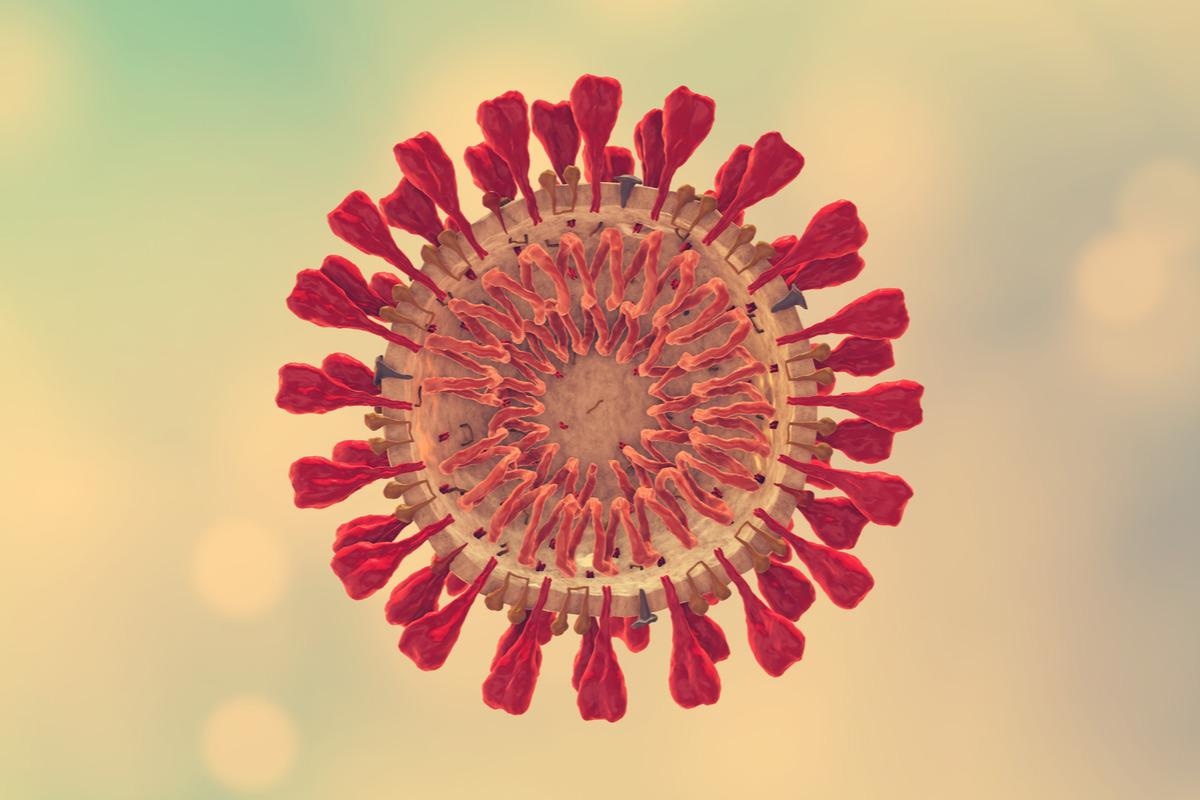
Research: SARS-CoV-2 spike protein–induced cell fusion prompts the cGAS-STING pathway and the interferon response. Picture Credit score: Cinefootage Visuals / Shutterstock.com
Background
A earlier research on people who died of COVID-19 reported three hallmarks of vital SARS-CoV-2 an infection which included the presence of large lung thrombosis, in depth alveolar injury, and syncytial dysmorphic pneumocytes. Syncytia, which arises resulting from cell fusion, has additionally been reported in SARS-CoV-2–contaminated macaques and in vitro infections. Thus, cell fusion performs an vital function in SARS-CoV-2 pathogenicity.
The SARS-CoV-2 spike (S) protein is accountable for triggering cell fusion. Following cleavage of the S protein, it engages with the cell floor angiotensin-converting enzyme 2 (ACE2) receptor for the fusion of the viral membrane with the plasma membrane of the host cell. Thereafter, viral ribonucleic acid (RNA) is launched into the host cell cytoplasm for replication.
SARS-CoV-2 makes use of a number of mechanisms to suppress the manufacturing of interferons (IFNs) through the early phases of an infection. A number of research have proven that SARS-CoV-2 an infection leads to the manufacturing of low quantities of sort I IFNs and excessive quantities of inflammatory cytokines, whereas others have proven that this an infection may also result in the manufacturing of excessive quantities of IFNs. This means that cells can sense SARS-CoV-2 an infection and mount an IFN response, regardless of the virus evading sure innate immune pathways.
People contaminated with SARS-CoV-2 can profit from safety by IFNs, particularly sort I and sort III IFNs. Nonetheless, extended manufacturing of IFN can result in impairment of lung epithelial regeneration.
The manufacturing of sort I and sort III IFNs is induced by the popularity of pathogen-associated molecular patterns by sample recognition receptors (PRRs). As an RNA virus, SARS-CoV-2 is detected by viral RNA sensors melanoma differentiation-associated protein 5 (MDA5) and retinoic acid-inducible gene I (RIG-I) in contaminated cells.
A brand new Science Signaling research analyzes how the activation of DNA sensor protein cytosolic cyclic GMP–AMP synthase (cGAS) and its downstream effector stimulator of interferon genes (STING) can induce a sort I IFN response resulting from SARS-CoV-2 an infection.
In regards to the research
The present research concerned S protein-mediated cell fusion assays, the place goal cells have been loaded onto the donor cells in a 1:1 ratio. Thereafter, S-protein mediated cell fusion and IFN manufacturing have been inhibited utilizing inhibitory brokers such because the furin inhibitor decanoyl-RVKR-chloromethyl ketone (RVKR) or the lysosomotropic inhibitor bafilomycin A1 (BaflA1).
Sequencing was carried out utilizing purified viral RNA adopted by RNA-seq knowledge evaluation and reverse transcription-quantitative polymerase chain response (RT-qPCR) evaluation. This was adopted by reporter assays, immunofluorescence evaluation, western blotting evaluation and imaging, in addition to 2′3′-cGAMP enzyme-linked immunosorbent assay (ELISA). Replication-competent vesicular stomatitis virus (VSV)–SARS-CoV-2 was used to verify the impact of S protein expression in fused cells.
Research findings
The S protein was discovered to change mobile transcriptomes and induce the transcription of IFNβ and tumor necrosis issue α (TNF-α), in addition to IFN-stimulated genes (ISGs) together with ACE2 and IFN-induced protein with tetratricopeptide repeats 2 (IFIT2) in fused cells. The S protein was additionally discovered to extend phosphorylation of the p65 subunit of nuclear issue κB (NF-κB).
Moreover, a rise within the nuclear translocation of the transcription issue IFN regulatory issue 3 (IRF3) was reported, together with a smaller ACE2 after cell fusion. The S protein was discovered to induce the sort I IFN response in a time-dependent method.
S protein mutants with an altered S1/S2 website or an altered S2′ website have been proof against cleavage by proteases. Nonetheless, the SARS-CoV S protein and S mutants with an altered S1/S2 website might generate modest syncytia, whereas mutants with an altered S2′ website have been unable to induce fusion. Notably, the activation of innate immune responses couldn’t happen with the 2 S mutants.
Stimulating the expression of IFNβ and the phosphorylation of IRF3 and STING required the expression of each cGAS and STING. The S protein was discovered to extend the manufacturing of two′3′-cGAMP in cells expressing cGAS and STING.
Knockdown of endogenous cGAS or STING was discovered to lower IFN expression, in addition to the phosphorylation of IRF3 and STING. Moreover, the activation of cGAS-STING was depending on cleavage of the S protein and didn’t happen in S mutants the place cleavage can not happen.
Cell-cell fusion led to elevated expression of 429 genes and decreased expression of 382 genes. A lot of the elevated genes have been related to cell senescence and DNA injury responses (DDRs). Moreover, the presence of micronuclei was noticed in S-induced fusion cells however not in fusion cells expressing mutant S proteins.
Notably, cGAS was localized within the micronuclei in fused cells and was related to the activation of IRF3. S protein expression and syncytia formation of contaminated cells was confirmed utilizing VSV–SARS-CoV-2, which carried the SARS-CoV-2 S protein. Moreover, cGAS activation and DNA injury have been noticed throughout an infection with VSV-SARS-CoV-2.
Conclusions
Taken collectively, the present research means that an infection with SARS-CoV-2 results in the activation of cGAS and STING resulting from micronuclei formation within the fused contaminated cell. Each cGAS and STING can subsequently activate IFNs and cytokines, which worsens illness severity by ultimately inflicting injury to the lung epithelial cells.
[ad_2]


